1.2 - Microscopy
Light microscopes
- A microscope lets us see things too small to see with the naked eye, which is why microscopes are central to studying cells.
- The light microscope is the older, more common type. It uses light and a system of lenses to form a magnified image of a specimen.
- Light microscopes were first used in the 1590s.
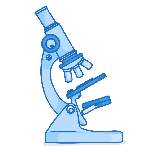
Key features
- Magnification - Up to roughly 1,500 times - enough to see individual cells and larger sub-cellular structures inside them, such as the nucleus and chloroplasts.
- Living specimens - Can be used to view living specimens, so cells can be studied while they are still alive.
Electron microscopes
- An electron microscope forms an image using a beam of electrons instead of light.
- They were developed in the 1930s.
- They have both a much higher magnification and a much higher resolving power than a light microscope - so they can show far smaller structures in far greater detail.
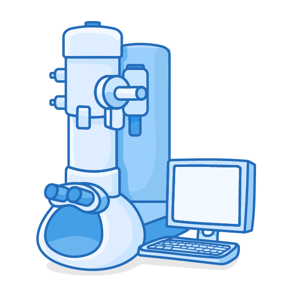
Revealing the inside of the cell
- Electron microscopes let biologists see the internal structure of cells for the first time, including:
- The inside of mitochondria and chloroplasts
- Very small features such as plasmids and viruses, where a light microscope showed nothing
- Seeing this internal detail let scientists work out how each structure carries out its job - for example how the inside of a mitochondrion is suited to respiration and how the inside of a chloroplast is suited to photosynthesis.
- The development of electron microscopy therefore greatly increased our understanding of how cells are built and how they work.
Drawbacks
- Expensive and not portable.
- Need a complicated process to prepare the specimen.
- That preparation kills the specimen, so - unlike a light microscope - an electron microscope cannot be used to view living material.
Comparing light and electron microscopes
- The two types of microscope suit different jobs.
- A light microscope is cheap, portable, and can view living specimens - but its lower magnification and resolution mean it shows only cells and their larger internal structures.
- An electron microscope has far higher magnification and resolution, revealing fine sub-cellular detail - but it is expensive, not portable, and cannot be used on living material.
Light vs electron microscopes
| Feature | Light microscope | Electron microscope |
|---|---|---|
| How it forms an image | Light and a system of lenses | A beam of electrons |
| First used | 1590s | 1930s |
| Magnification | Up to about x1,500 | Much higher (up to around x500,000) |
| Resolution (detail) | Lower | Much higher |
| What it can show | Whole cells and large structures such as the nucleus and chloroplasts | The internal structure of mitochondria and chloroplasts, plasmids, viruses |
| Living specimens | Can be viewed | Cannot (specimen is killed in preparation) |
| Cost and portability | Cheap and portable | Expensive and not portable |
TermDefinition
Light microscopeA microscope that uses light and a system of lenses to form a magnified image of a specimen.
Electron microscopeA microscope that forms an image using a beam of electrons instead of light.